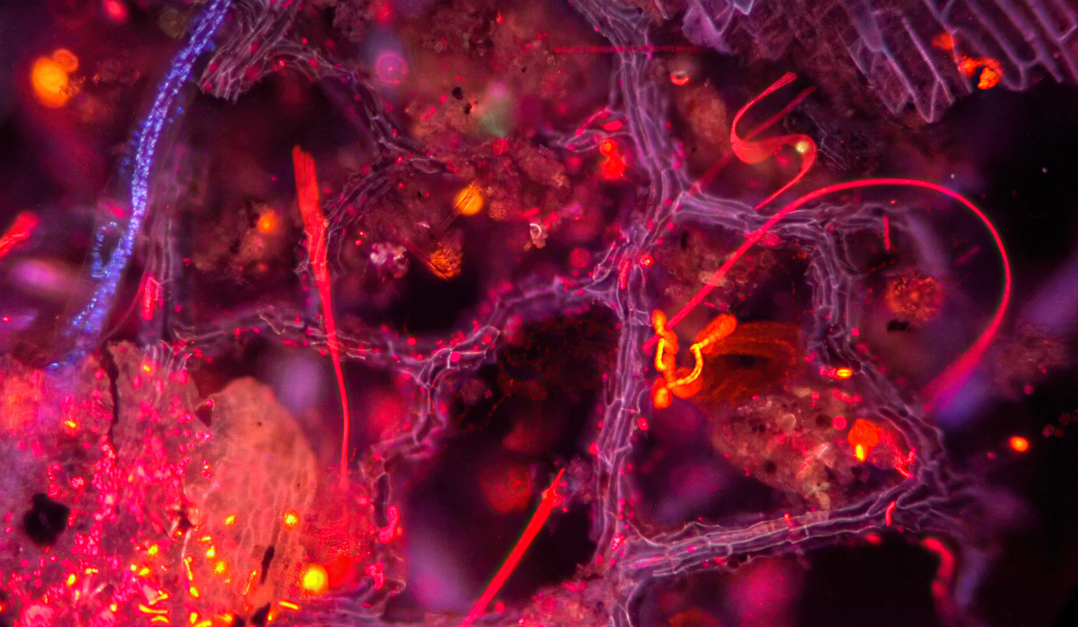
Fluorescence
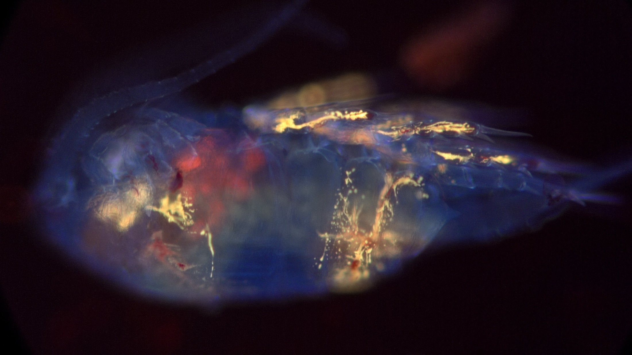
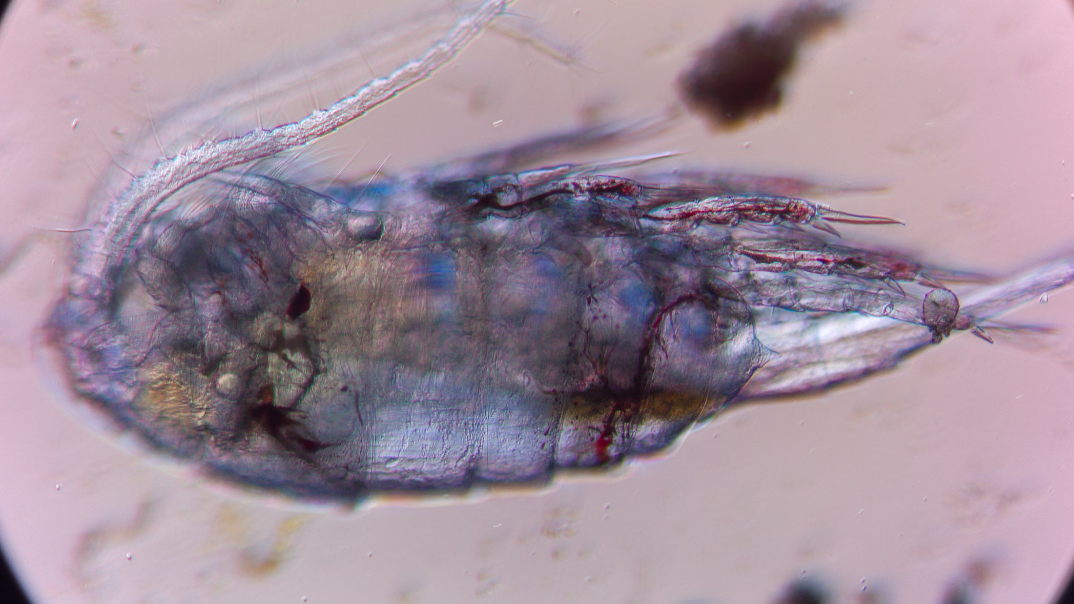
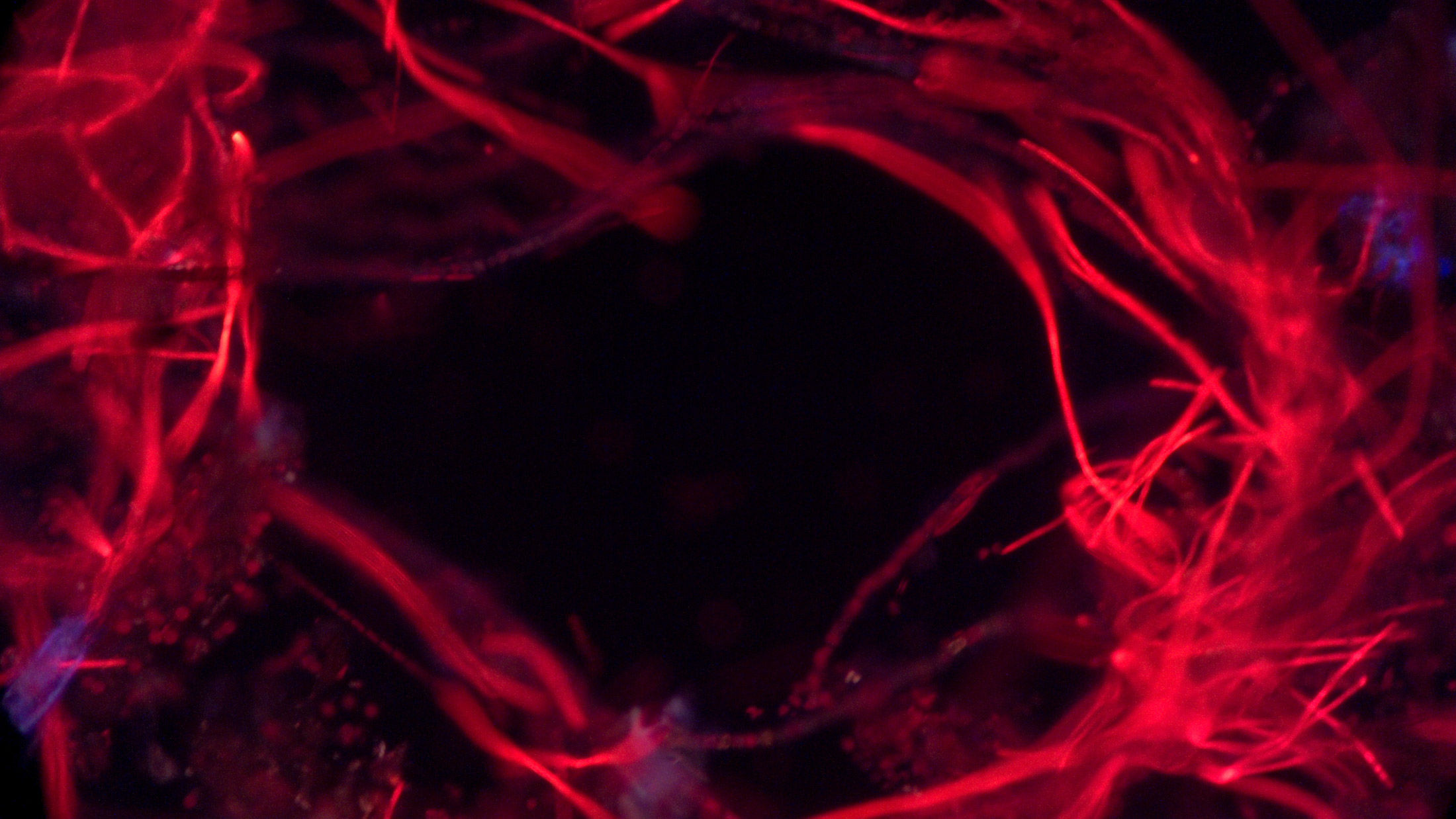
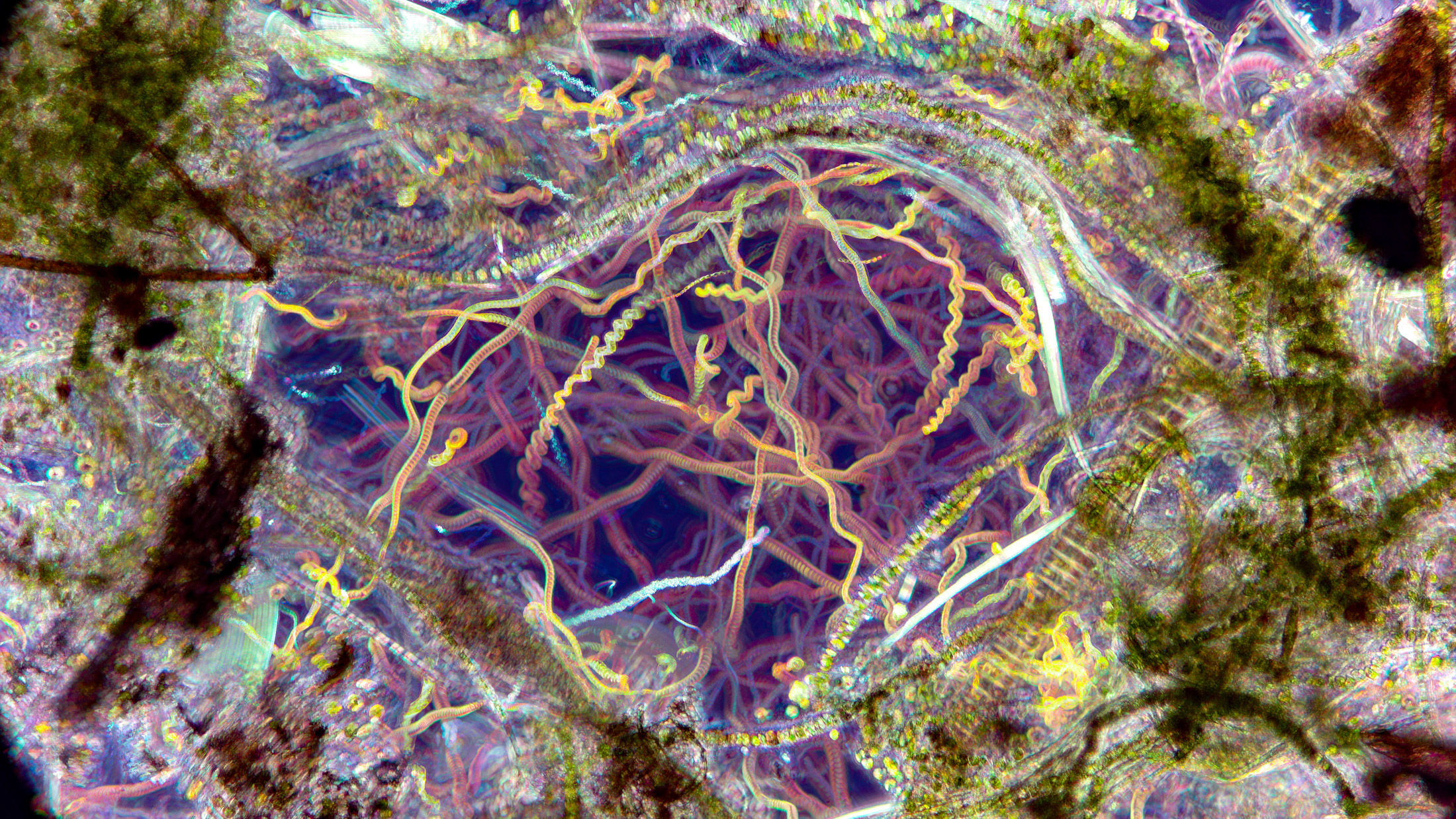
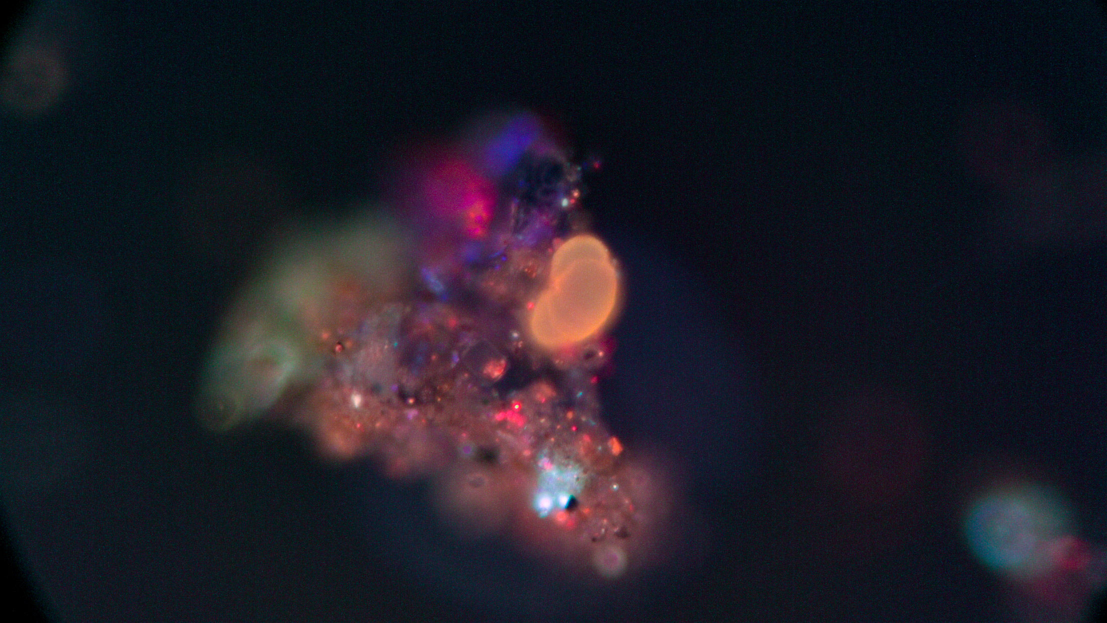
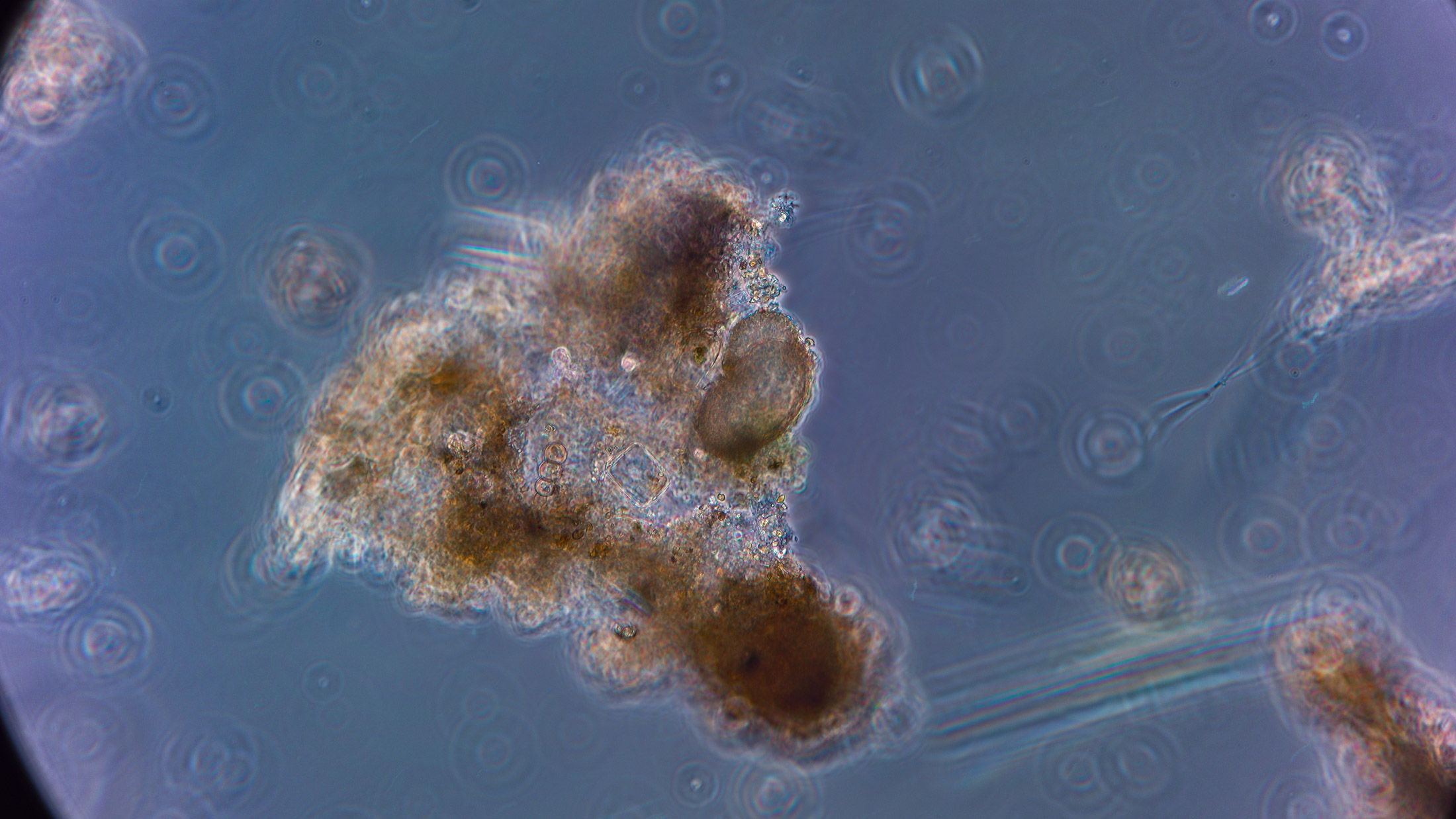
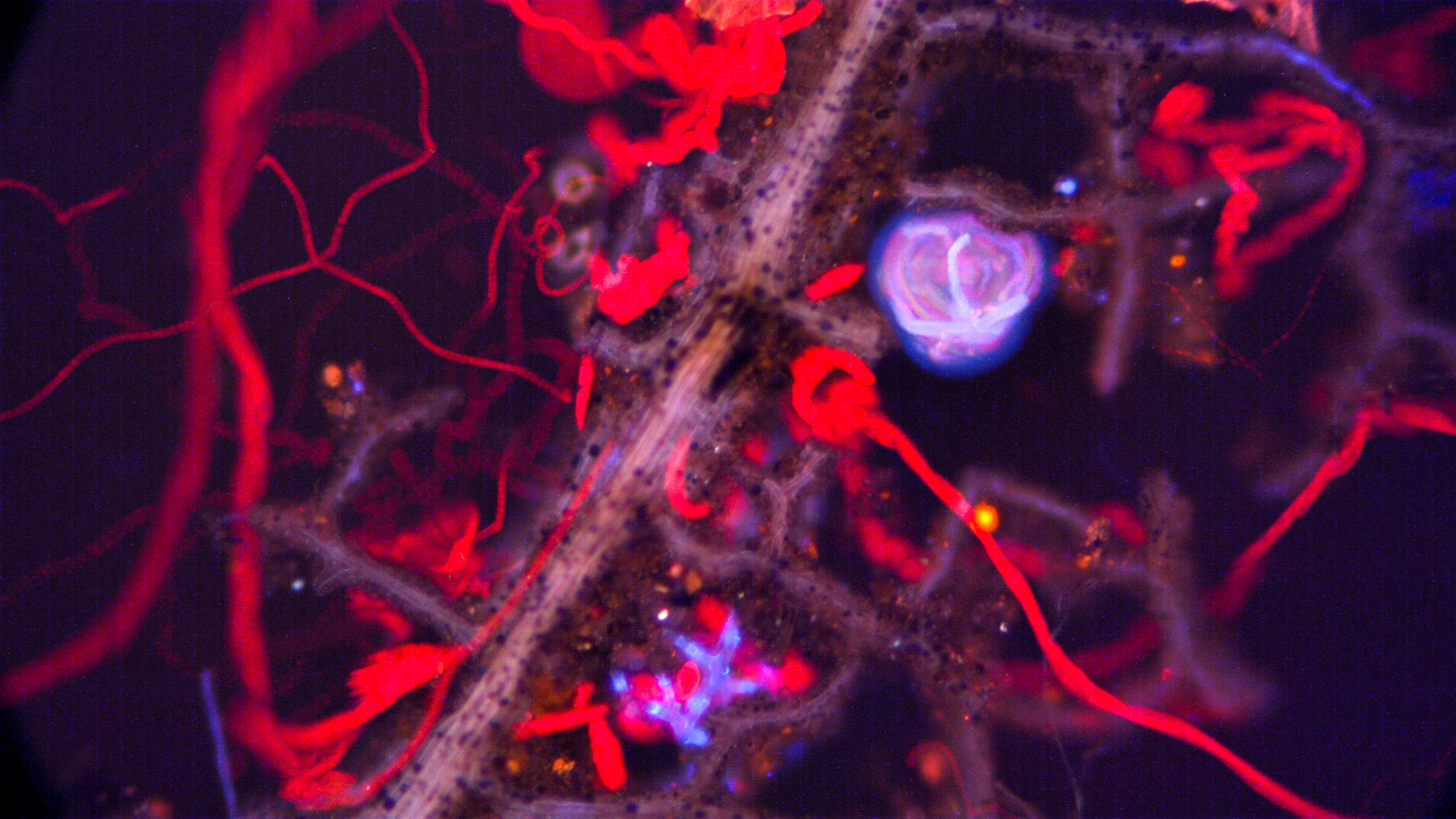
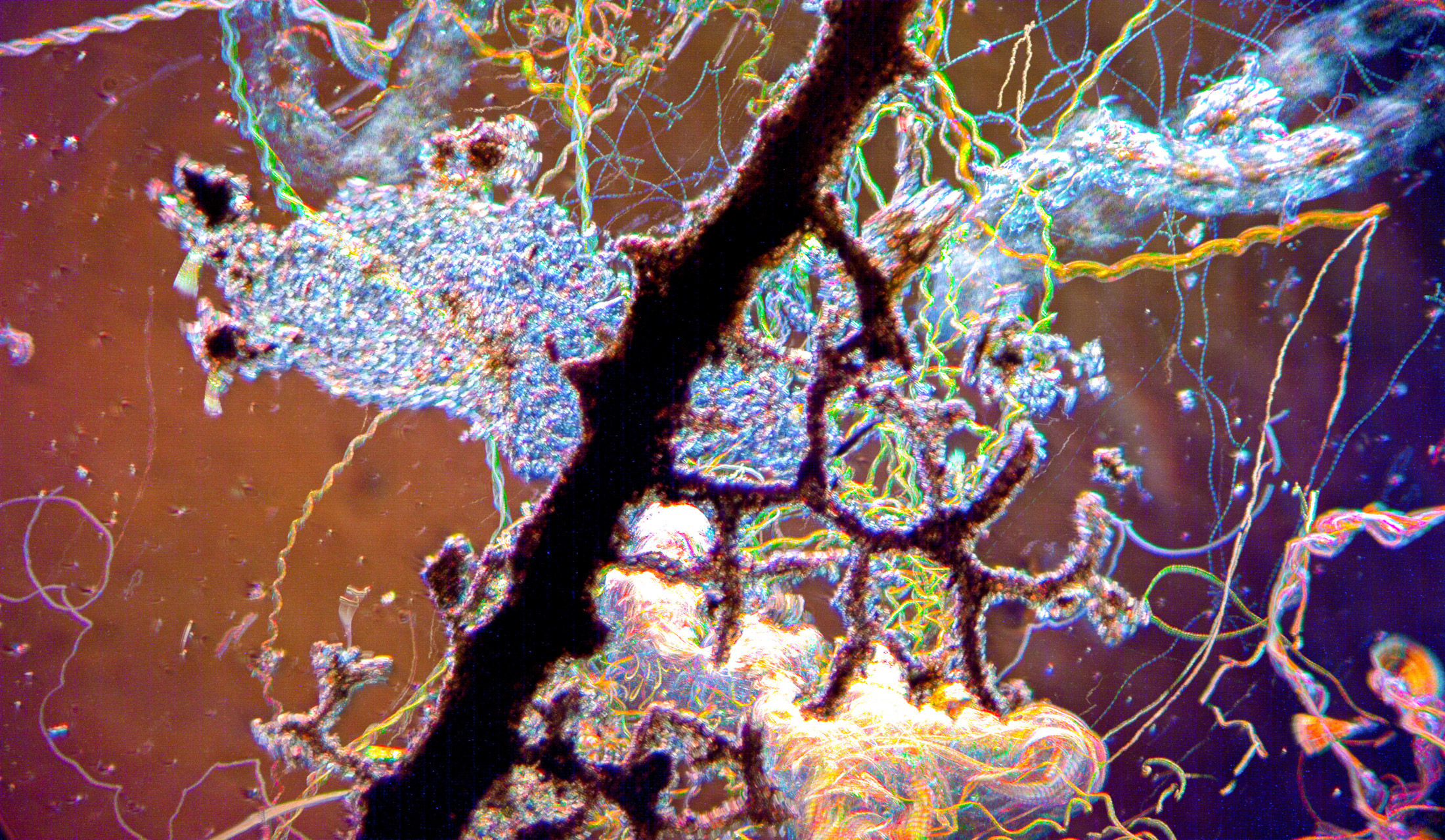
This series explores the natural autofluorescence of microscopic organisms, algae, plant material, and aquatic debris under fluorescence microscopy. By exciting samples with specific wavelengths of light, structures normally hidden in transmitted-light microscopy begin glowing against darkness in vivid reds, blues, violets, and magentas. Cell walls, chitinous shells, chlorophyll-rich tissues, and suspended particles emerge as luminous architectures and drifting signals, transforming familiar microscopic life into something strangely spectral and immersive. The work explores how fluorescence imaging reshapes perception, revealing a microscopic world that feels both biological and otherworldly.
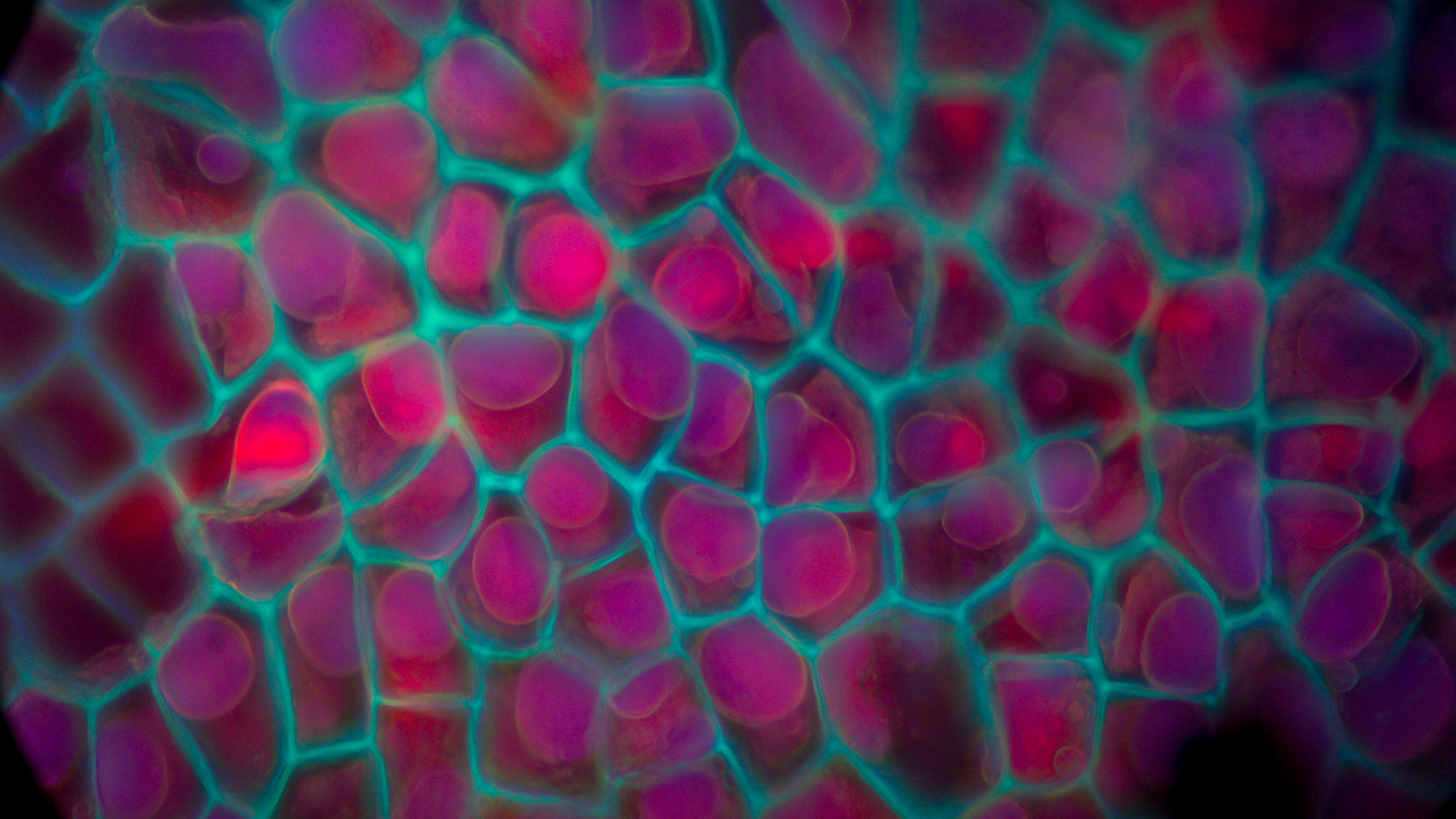
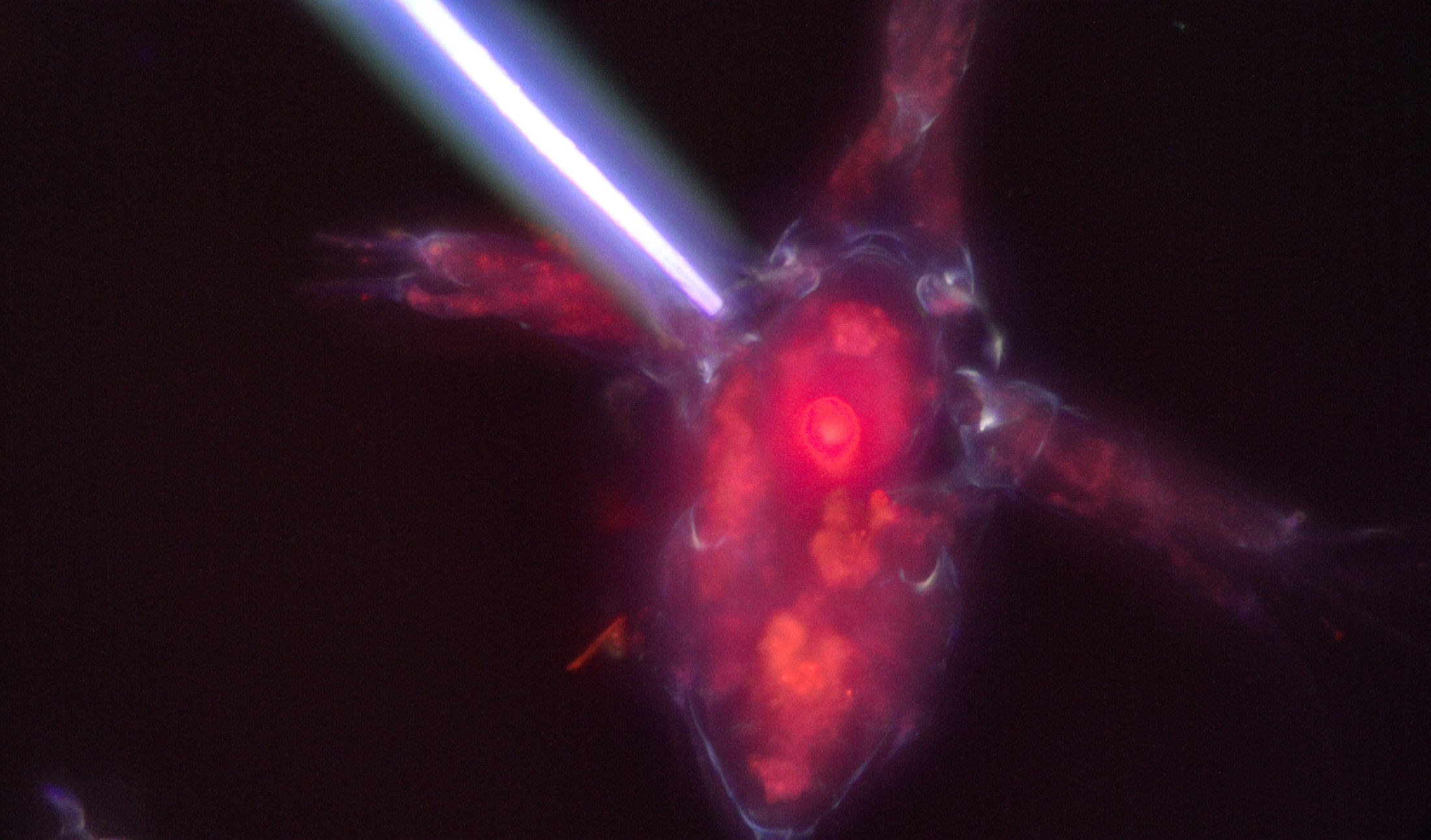
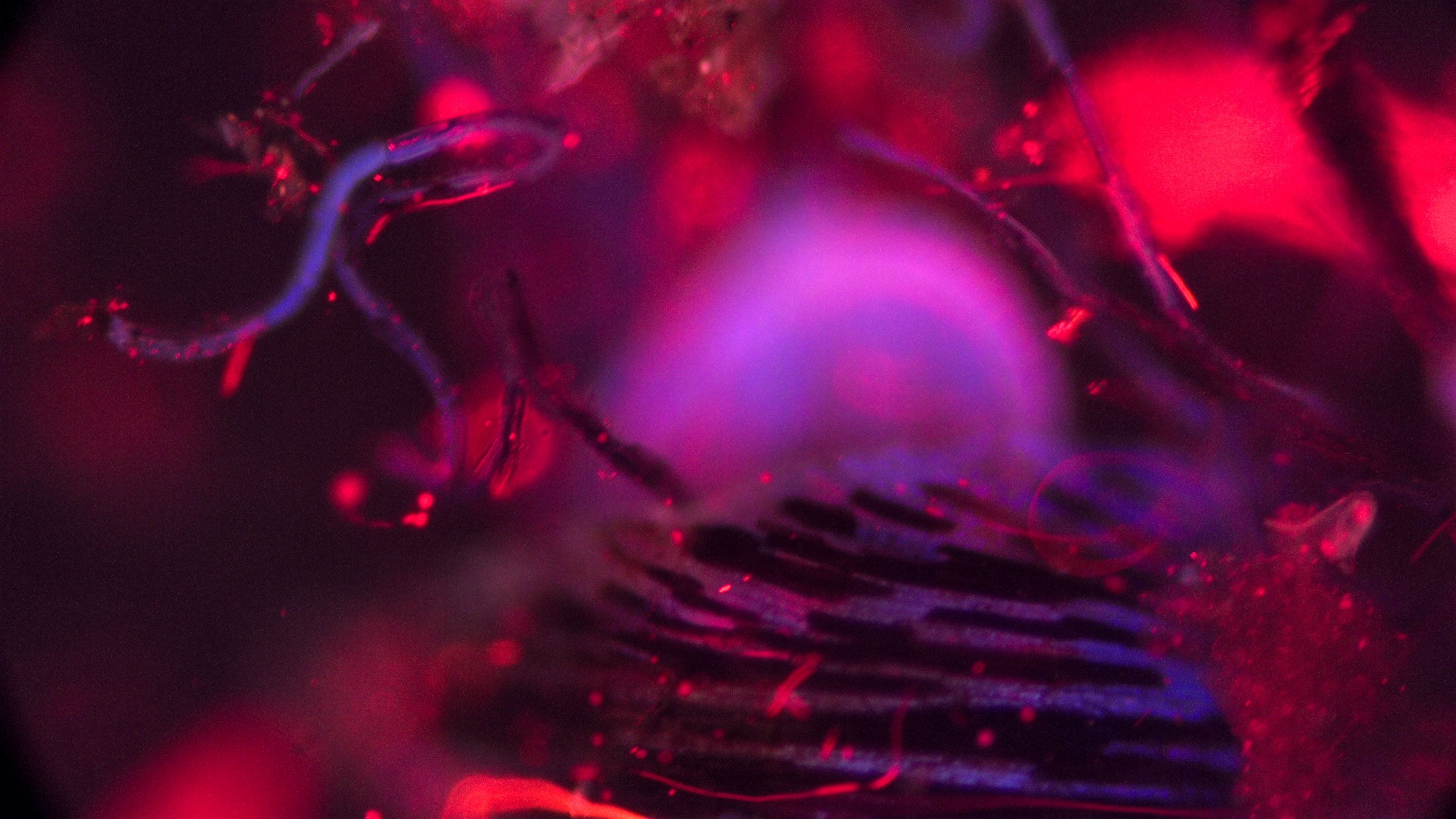
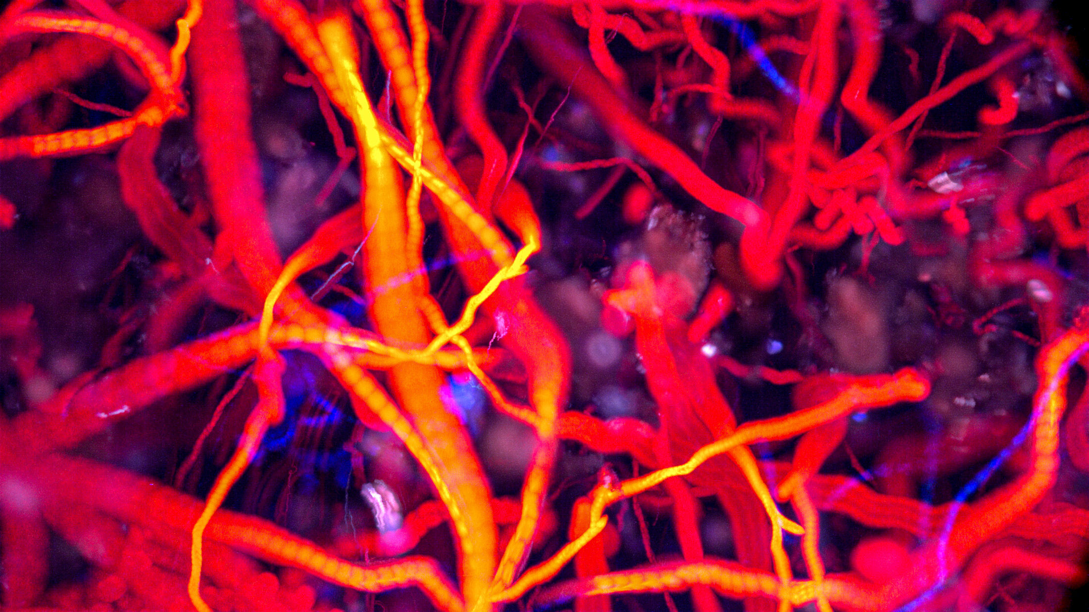
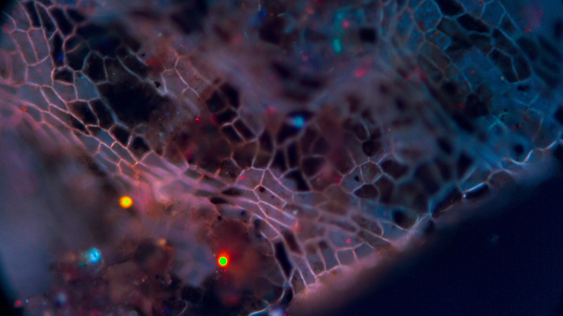
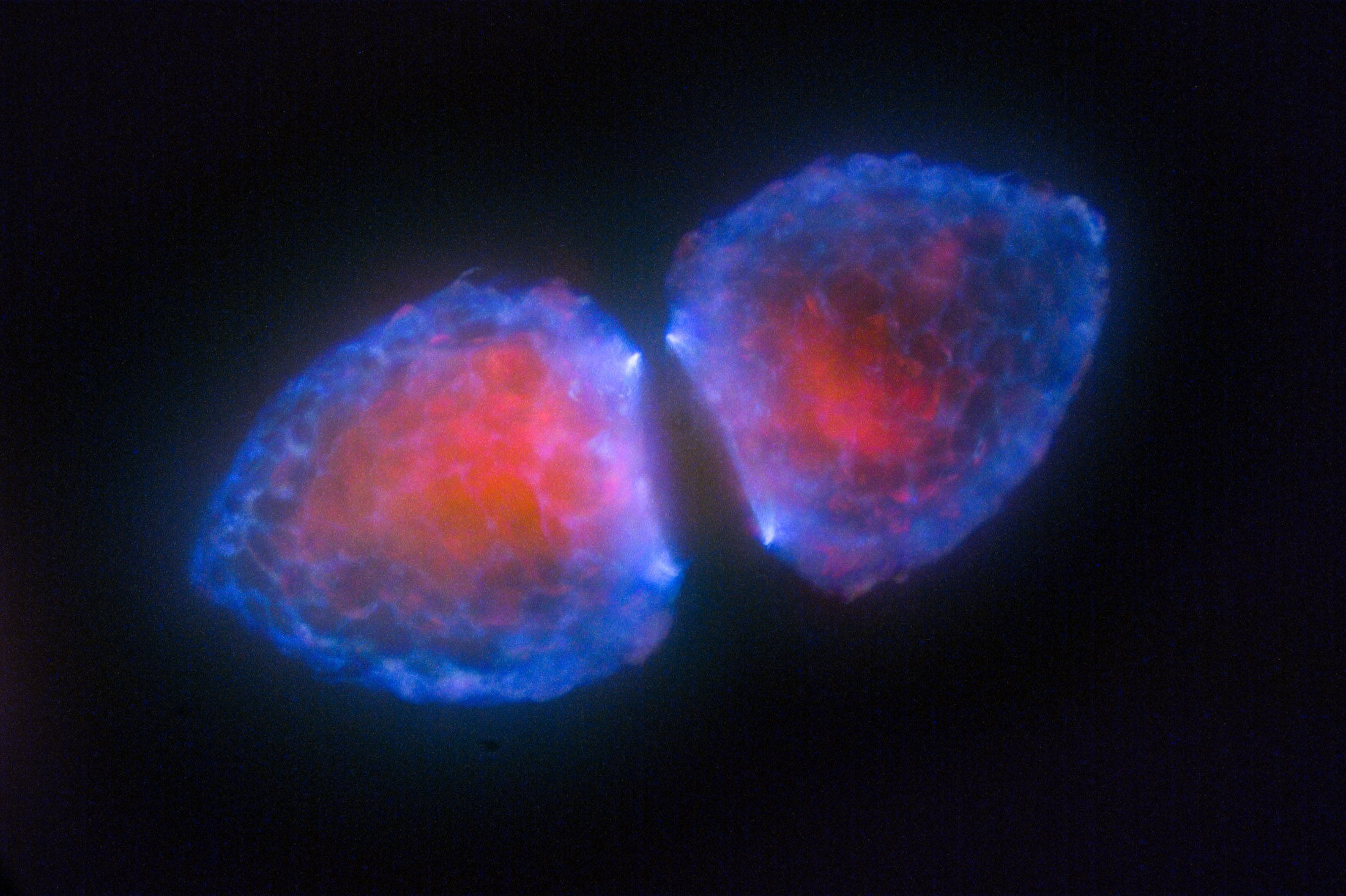
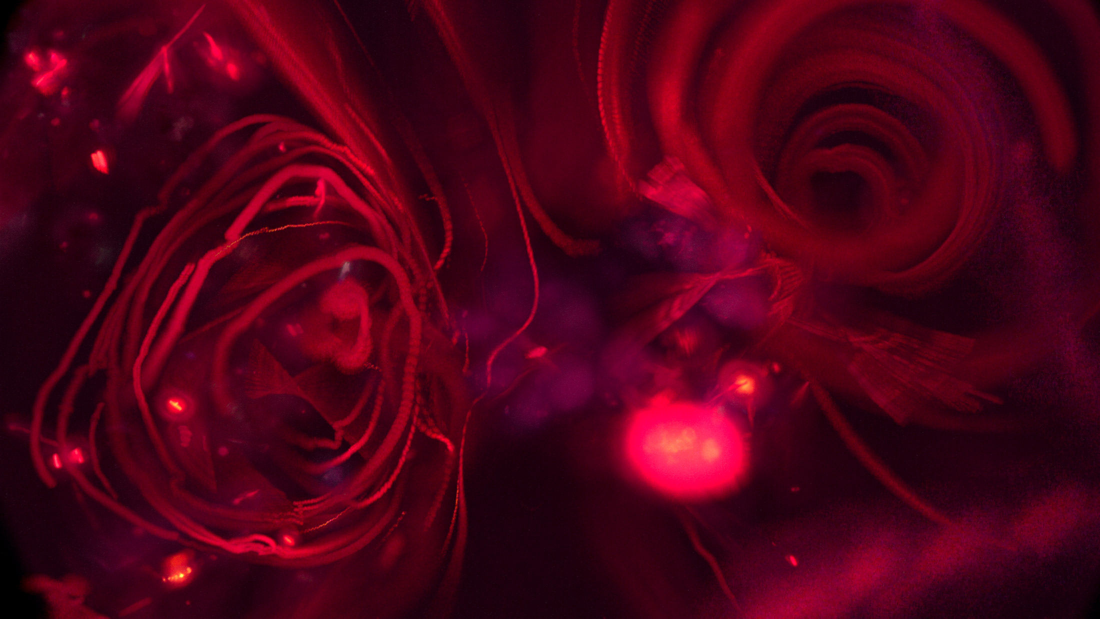
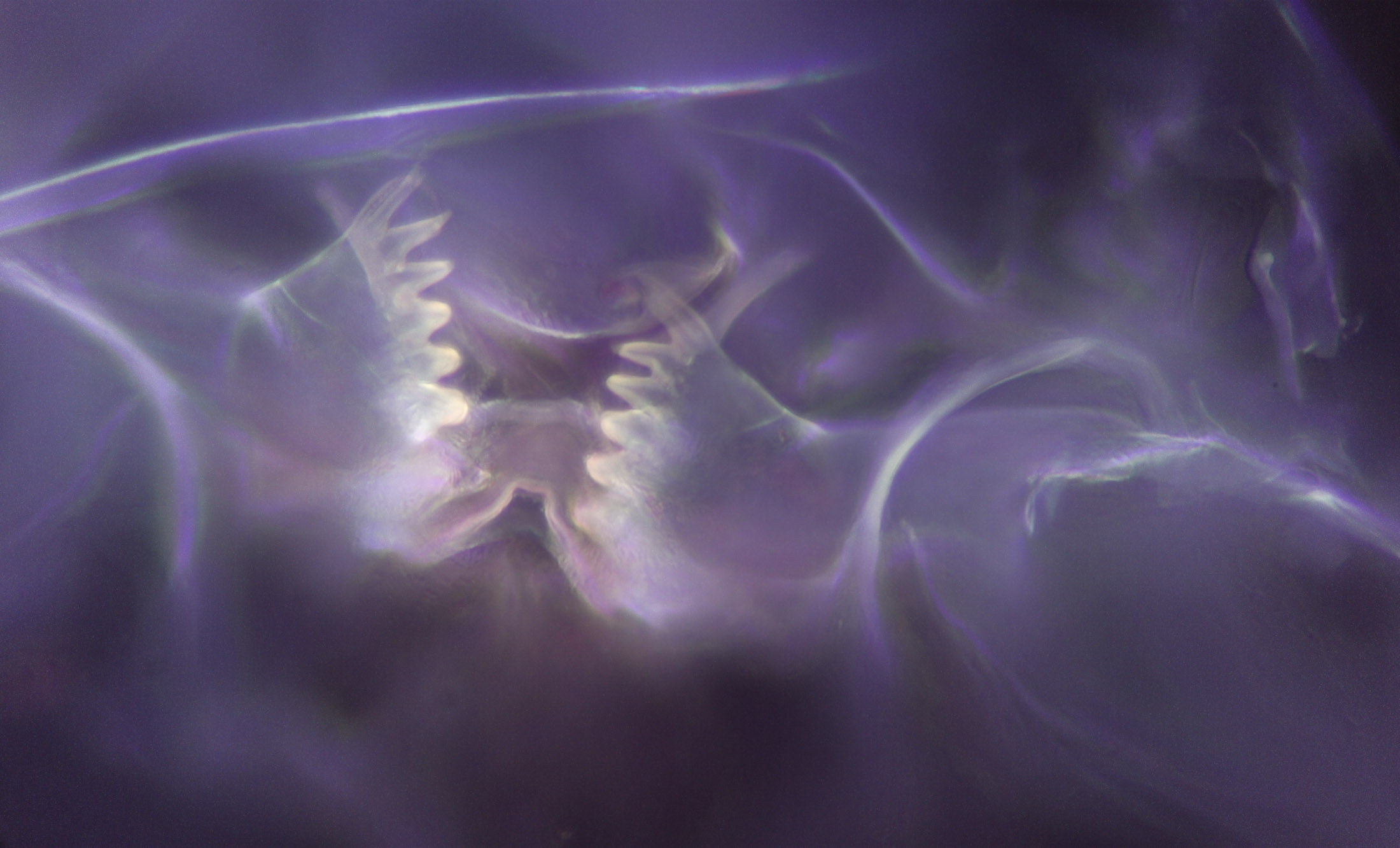
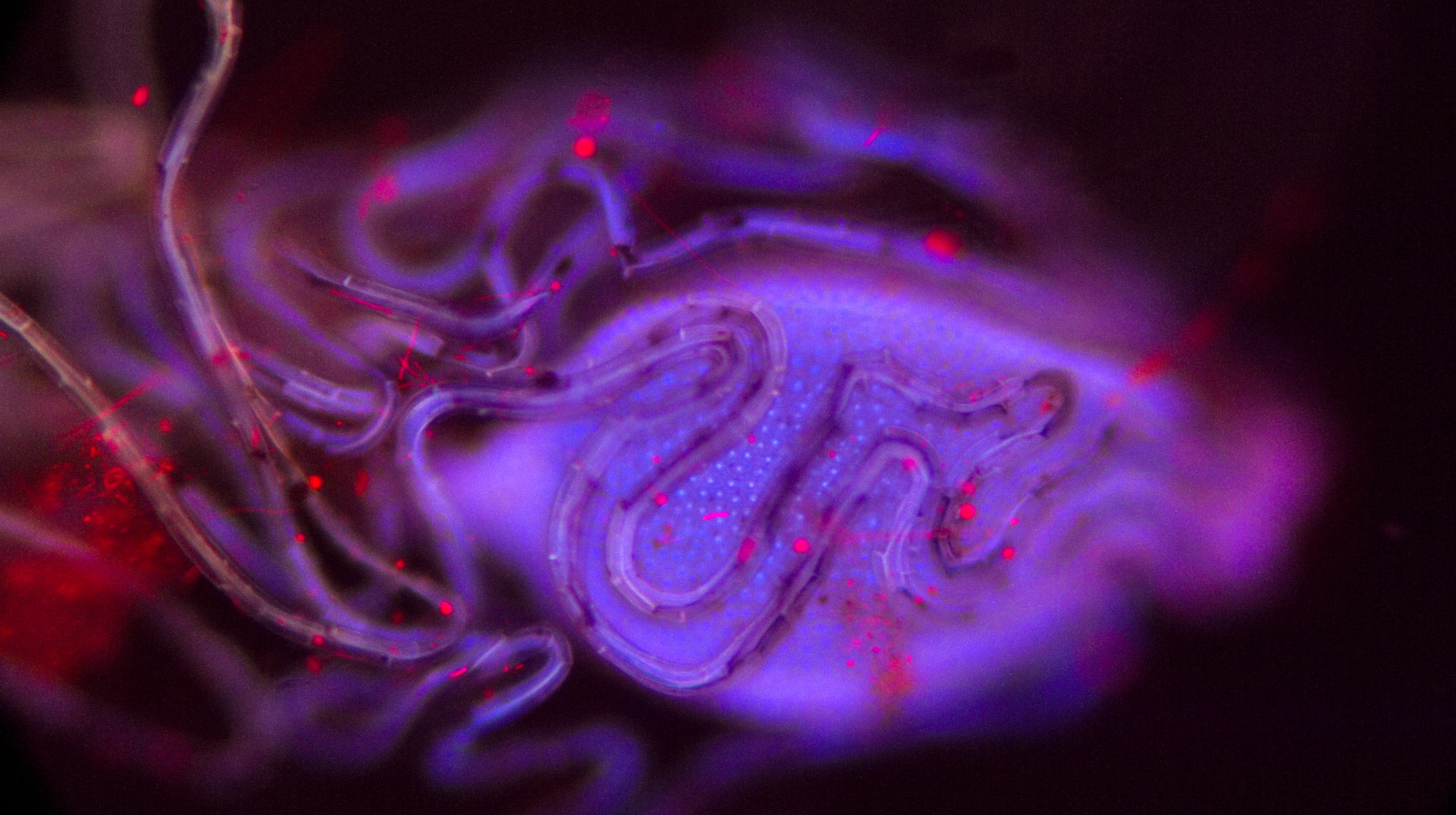
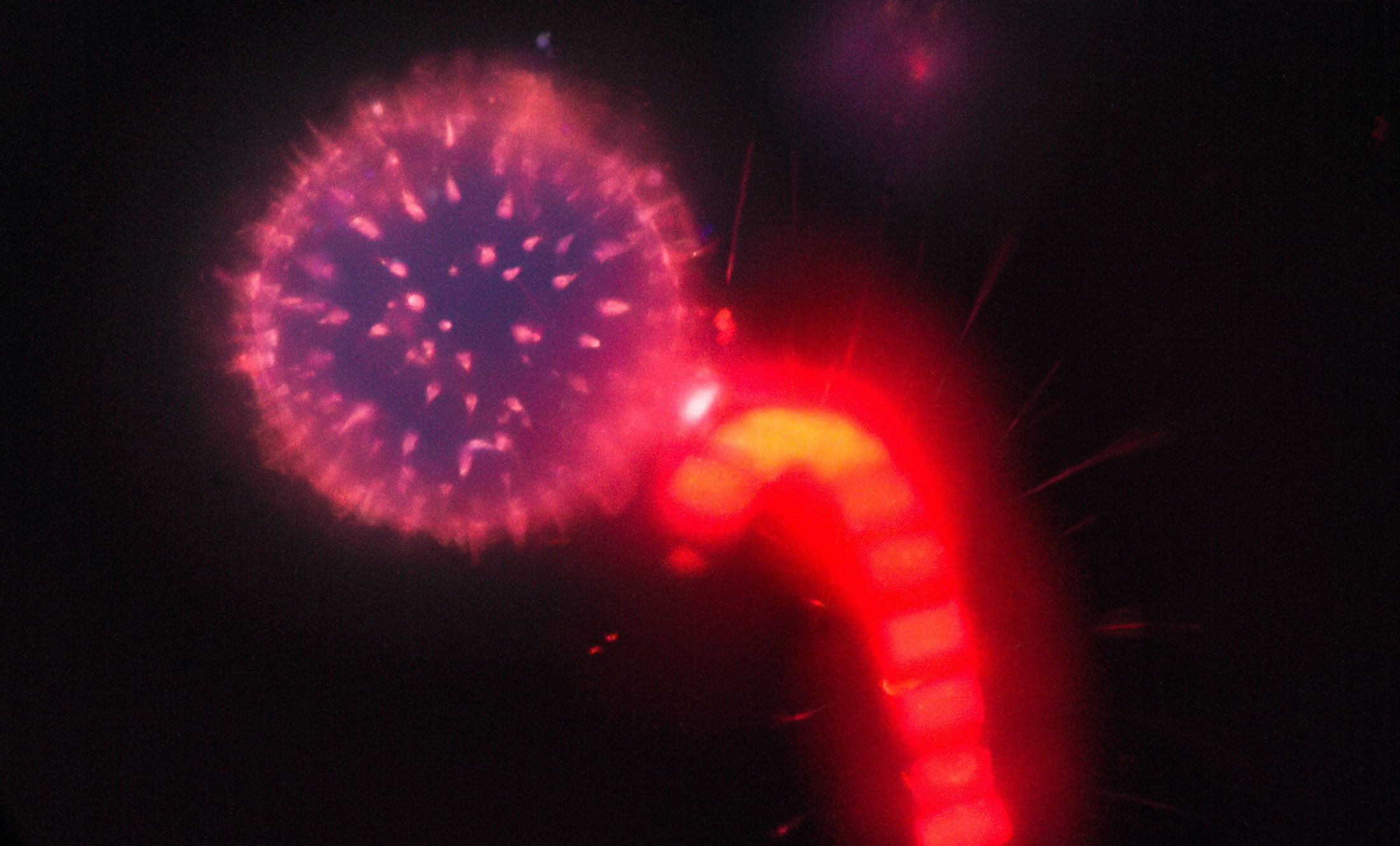
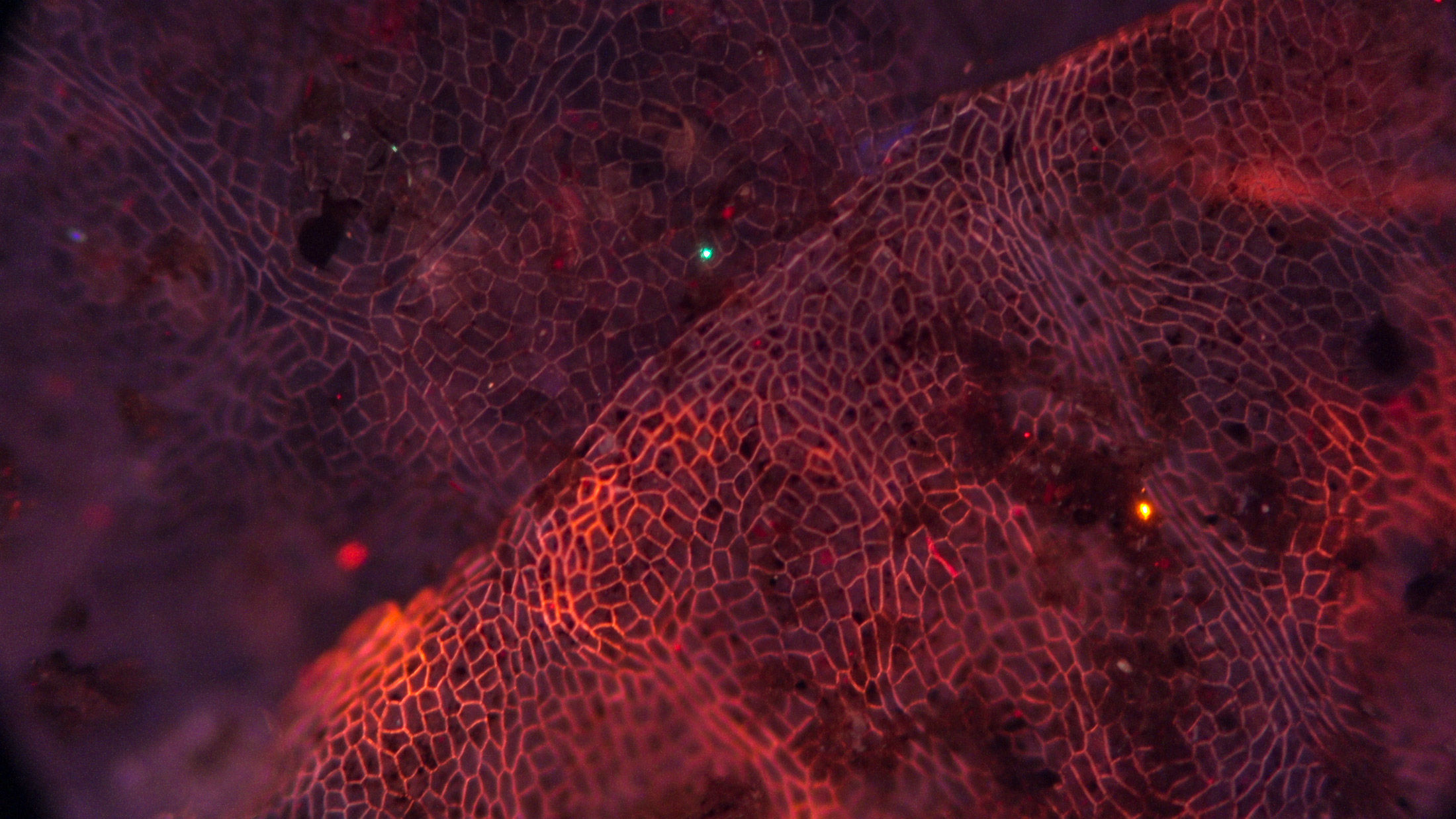
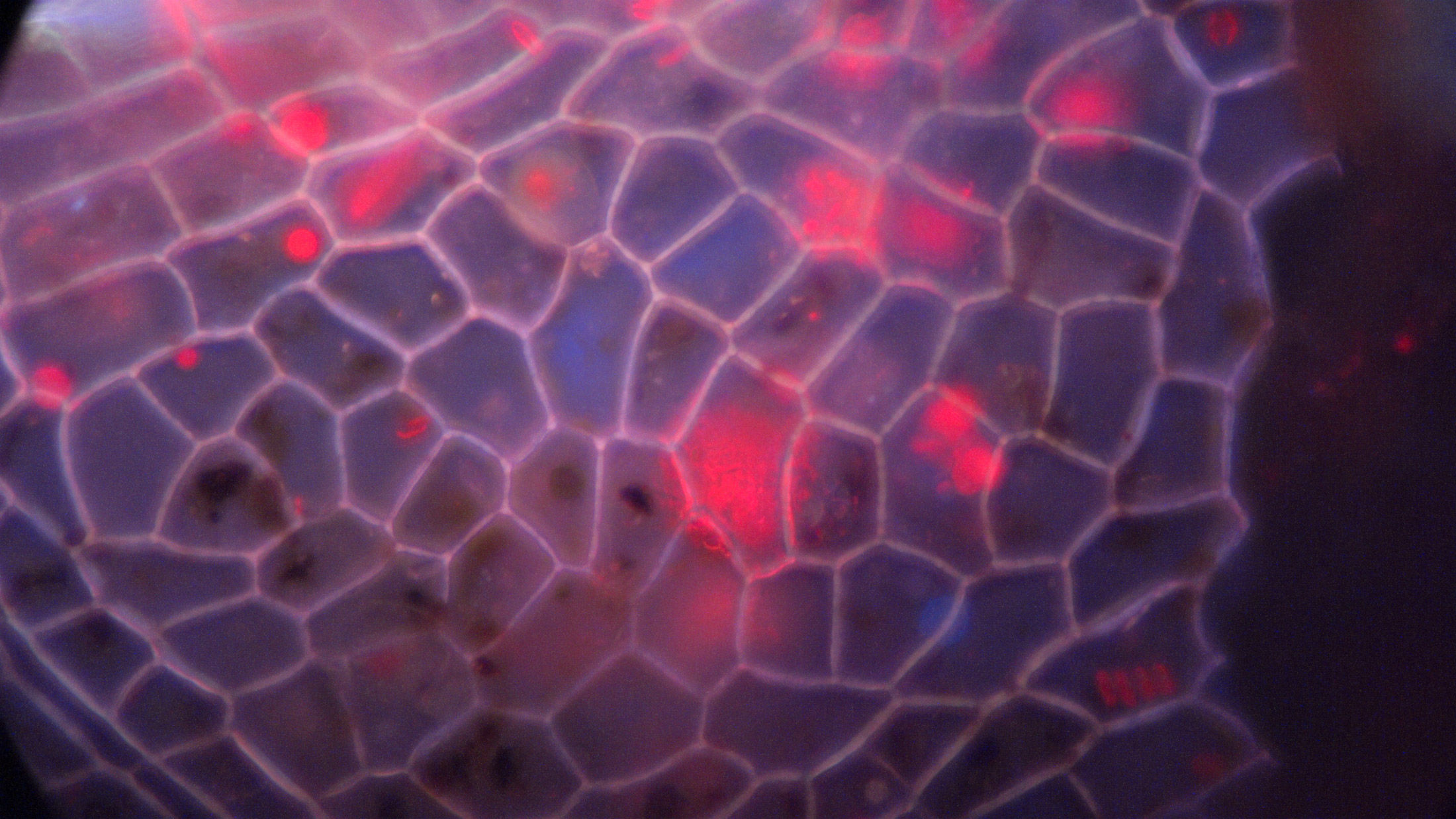
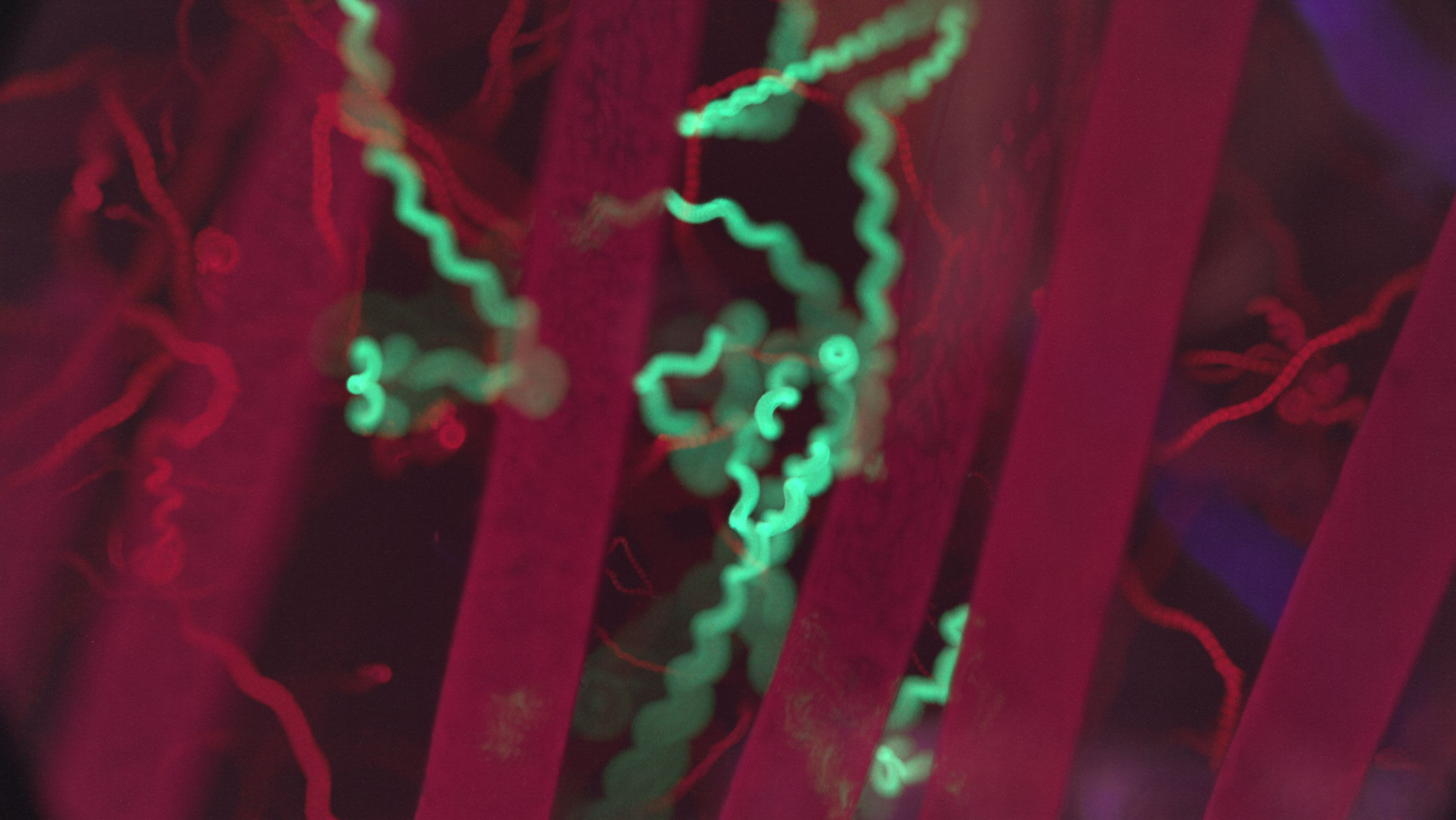
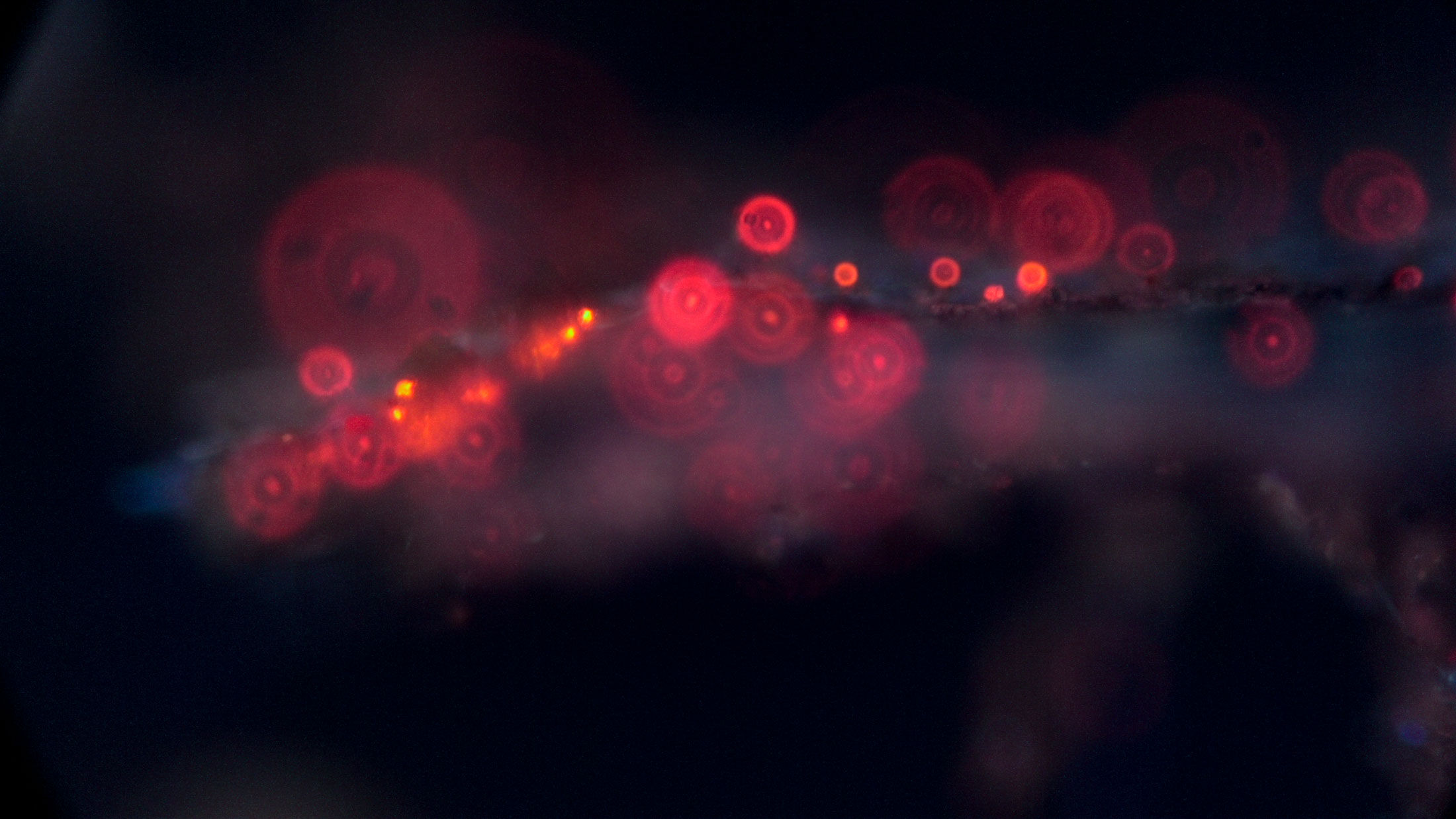
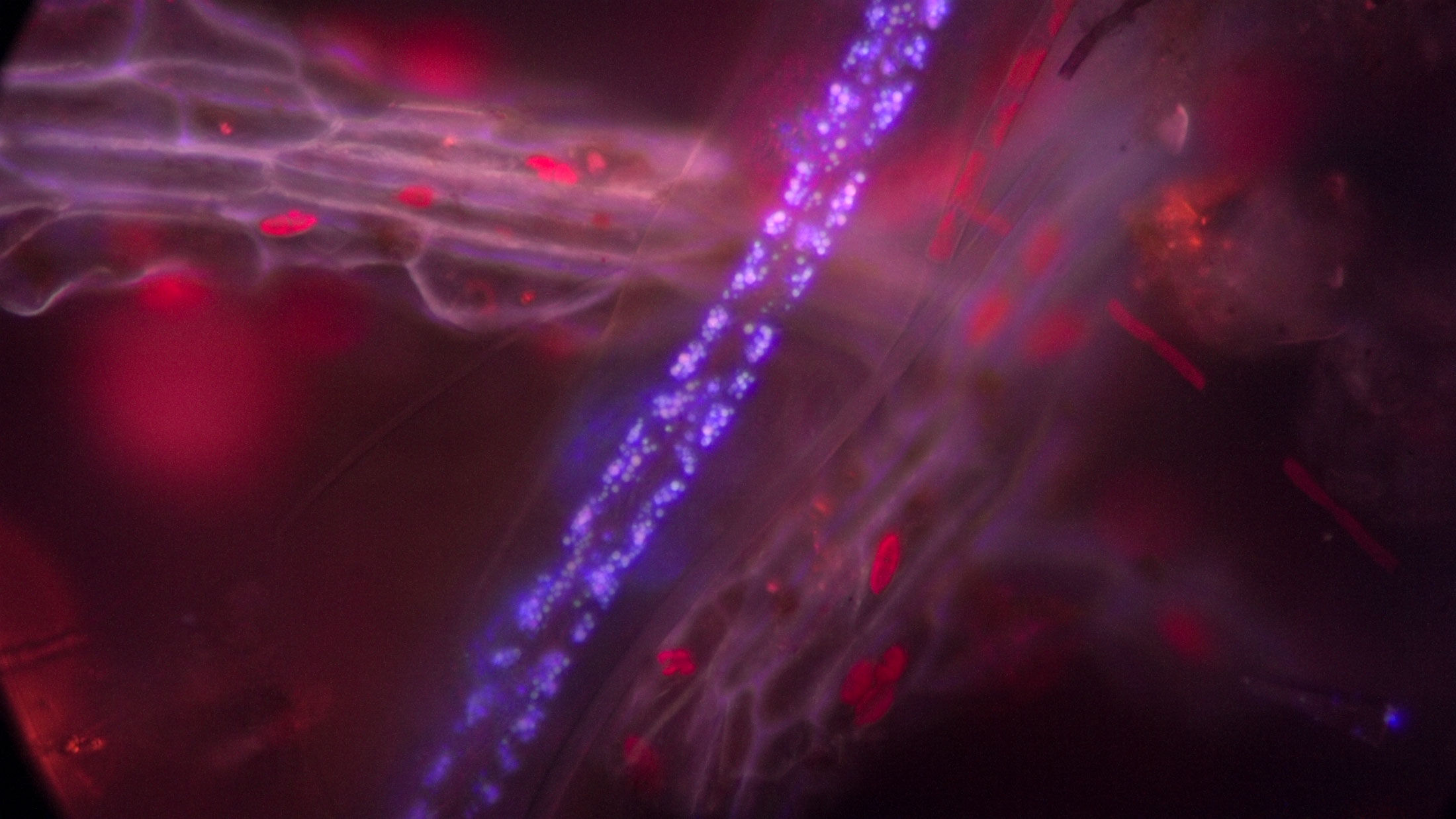
Excitation: 380/470/557nm SOLA light engine Emission: 428/511/627nm triple bandpass Exposure: 16-60ms Camera: Sony RX0 II
Fluorescence microscopy works by illuminating specimens with specific wavelengths of light that excite naturally fluorescent molecules within the sample. Chlorophyll strongly fluoresces deep red, while chitinous structures often glow blue or cyan. Algae, microorganisms, plant tissues, and suspended debris produce fluorescent signatures that reveal chemistry and structures largely invisible under traditional transmitted-light methods. Against the dark background, organisms often appear suspended or disembodied, giving the microscopic world an unusually haunting presence.